Home -> Vectors -> Ticks -> Morphology
Morphology
A characteristic of the Acarines is the extreme fusion of body segments, in contrast to the known three body segments head, thorax and abdomen in insects.
The integument
The tick’s integument is composed of the epidermis and the cuticle. The cuticle is the dead, outer part of the integument. It is secreted by the epidermis which forms the inner, living part of the integument.
The cuticle can be differentiated into the thinner epicuticle and the much thicker procuticle of protein and chitin. However, in ixodid ticks the outer part of the procuticle, termed exocuticle, becomes sclerotised in certain regions, i. e., the scutum.
The integument serves as a body covering, as the primary protection against water loss and also as the exoskeleton. It provides protection against mechanical and other types of physical damage.
The nervous system
No part of the central nervous system is located within the gnathosoma of the tick, which therefore does not correspond to the head in the generalised arthropod. The tick central nervous system is more condensed than in other Chelicerata.
The "brain", termed synganglion, is located centrally at the level of the second coxae.
The synganglion is formed by the fusion of the brain ganglia and the abdominal nerve cord into a single mass. As in other acari it is divided into two parts by the esophagus.
The cranial, preesophageal part consists of the protocerebrum, the optic lobes, the cheliceral and pedipalpal ganglia, and the stomodeal pons or bridge. All ticks examined have been found to possess well-developed photoreceptors, even the "eyeless" ticks (Aponomma, Ixodes, Haemaphysalis). They also have optic nerves and optic ganglia in the brain. A set of paired nerves extends from the optic lobes, a second set of paired nerves serves the chelicerae, and a third innervates the pedipalps. The unpaired stomodeal or pharyngeal nerve innervates the pharynx.
The postesophageal part of the synganglion gives rise to four pairs of pedal ganglia serving the four pairs of legs in the adult tick. Fine "sympathetic'' nerves connect all four pedal nerve trunks laterally on each side of the synganglion. Several pairs of opisthosomal nerves innervate the viscera. The ventral lobes of the pedal ganglia of leg 1 contain discrete areas of highly differentiated neuropile which are thought to receive olfactory fibres from pedal nerve 1 and have been called olfactory lobes.
Associative centers are represented by several bilaterally symmetrical glomerular structures.
Anterodorsal, posterodorsal, and ventral glomeruli in the pre-esophageal part are connected by nerve fibre trunks. A complex of nerve fibres and trunks in the postesophageal part of the synganglion forms a five-level commissure-connective system.
The synganglion and all peripheral nerves are covered by a connective tissue sheath, the neurilemma, below which there is a relatively thin layer of glial cells, the perineurium. Beneath these layers, subperineural glial cells are located on both sides of a cortex layer of nerve cell bodies surrounding the central fibrous neuropile, which constitutes the greater part of the synganglion mass (Mehlhorn, 2001).
The nervous system is closely associated with the circulation system. The entire central nervous system is enclosed within a perineural sinus of the circulatory system; this receives a dorsal aortic vessel and gives rise to vessels enclosing the major nerve trunks.
Locomotion
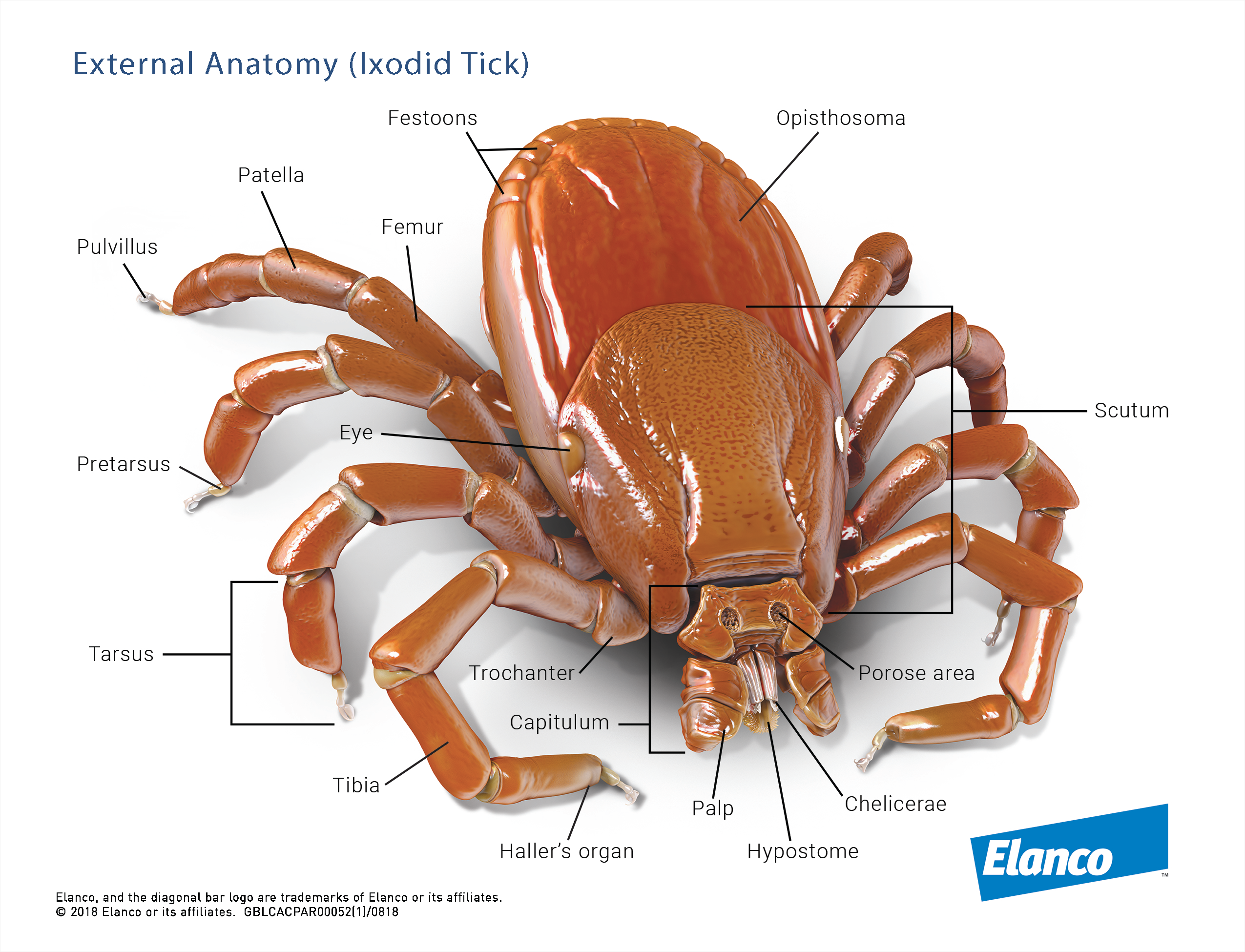
Ticks are typically acarine in having hexapod (six legs) larvae, and octapod (eight legs) nymphs and adults. Their legs are jointed and divided into seven segments: coxa, trochanter, femur, genu, tibia, tarsus and pretarsus.
The terminal pretarsus consists of a basal stalk, paired claws and a membraneous pulvillus. The pulvillus is absent in argasid ticks (Mehlhorn, 2001).
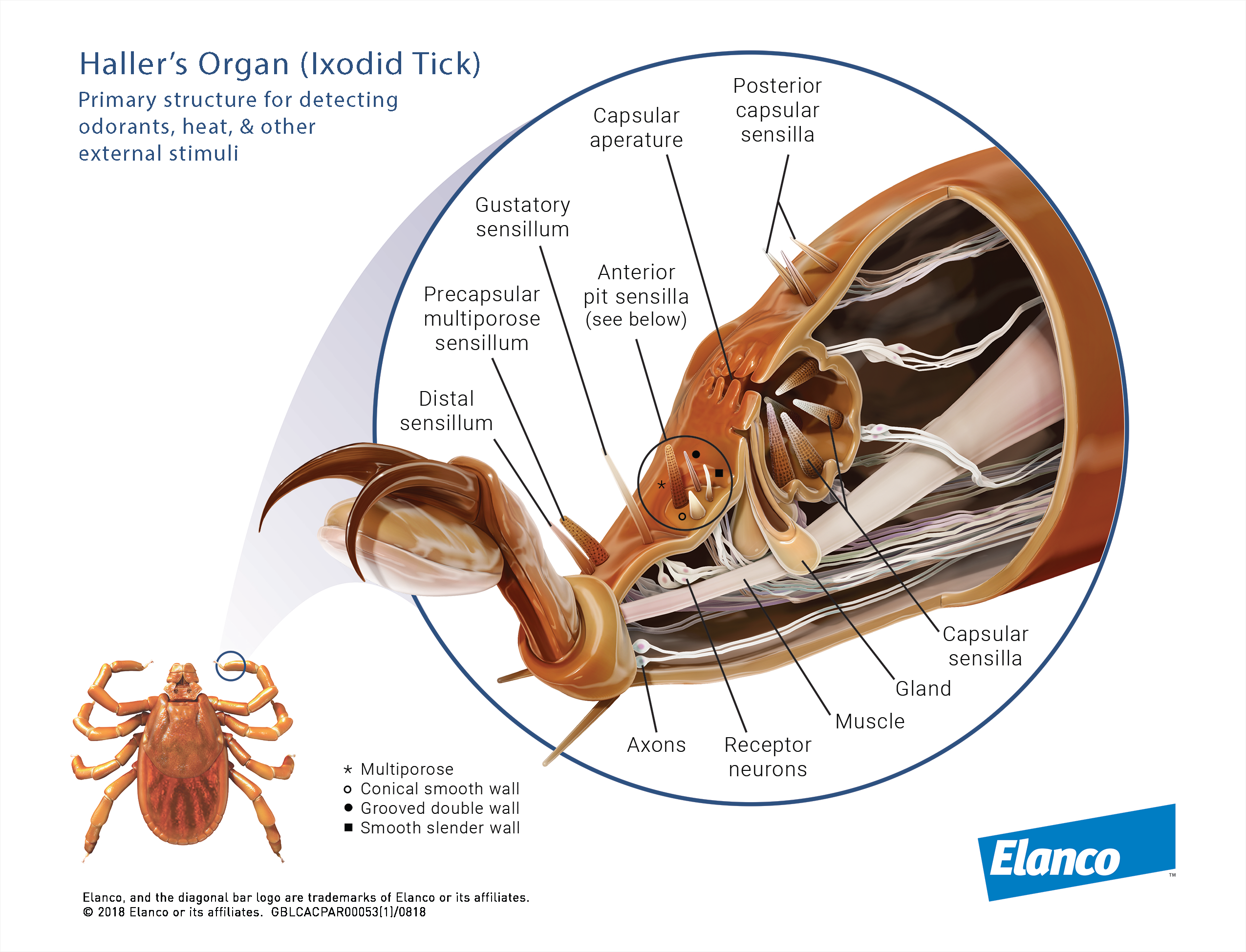
While the legs are primary ambulatory, they may be modified to serve other functions. The legs of Acari may be smooth or variously ornamented and usually possess a number of tactile and sensory hair (Krantz, 1978). On the dorsal surface of tarsus I, the Haller's organ, a complex sensory structure is found.
References
Krantz GW: A Manual of Acarology. 2nd edn., 1978, Oregon State Univ. Book Stores, Corvallis
Mehlhorn H: Ticks. In: Mehlhorn H (ed.): Encyclopaedic reference of parasitology. Vol. 1: Biology, structure, function. 2nd edn., 2001, Springer Verlag, Berlin, pp. 608-38
EXPLORE OUR CONTENT
 CVBD MapsThe CVBD Occurence World Map presents country-specific situations based on current scientific knowledge and feed-back from experts around the world in an easy-to-grasped way. |
| Read more-> |
 ResourcesElanco Animal Health supports education in parasitology and especially in the field of vector-borne diseases. Access image collections, discover the World Forum calendar, interesting links and our glossary. |
| Read more-> |
 CVBD World ForumThe CVBD World Forum is a working group of leading international experts with the mission to enhance knowledge and communication on companion animal vector-borne diseases for the improvement of animal, human, and environmental health. |
| Read more-> |